difference between rough endoplasmic reticulum and smooth endoplasmic reticulum
Thursday, December 28, 2023

Bell palsy vs facial palsy due to stroke
Bell palsy vs facial palsy due to stroke
For learning anatomy, please visit :
My youtube channel : @easyhumanatomy73
My website : http://easyhumananatomy.com
My facebook page: https://www.facebook.com/easyhumanatomy/
My blog: http://www.easyhumanatomy73.blogspot.com
My blog: Difference between : http://www.microscopicanatomybd.blogspot.com
The two most common causes of acute facial paralysis are Bell’s palsy and ischemic stroke or upper motor type of facial paralysis.
Facial weakness can be caused by strokes in many different locations in the brain and brainstem. Strokes involving the brain typically cause central facial weakness that involves the mouth and spares the eye and forehead.
Strokes involving the brainstem can sometimes cause weakness of the mouth, eye and forehead–mimicking a peripheral lesion. In these cases however, there will be other focal neurologic deficits. A review of systems and neurologic examination can help to identify signs and symptoms of stroke.
Bell's palsy is a condition that causes sudden weakness in the muscles on one side of the face. In most cases, the weakness is temporary and significantly improves over weeks.
The weakness makes half of the face appear to droop. Smiles are one-sided, and the eye on the affected side resists closing.
Bell palsy vs facial palsy due to stroke
Topic | Upper motor type/ stroke | Lower motor type/ bell’s palsy | |
Age | >60 years | 20 -50 | |
Time course | Second to minutes | Few hours to few days | |
Upper face | Usually not affected | Affected | |
Lower face | Affected | Affected | |
Associate symptoms | • Rapid onset of mild weakness to total paralysis on one side of your face — occurring within hours to days • Facial droop and difficulty making facial expressions, such as closing your eye or smiling • Drooling • Pain around the jaw or in or behind your ear on the affected side • Increased sensitivity to sound on the affected side • Headache • A loss of taste • Changes in the amount of tears and saliva you produce | stroke causing isolated left lower facial weakness. There’s a flattened nasolabial fold & inability to smile on the affected side with sparing of the forehead & eye closure muscles. Weakness or numbness in the arm or leg: Weakness or numbness can occur either on the same side as the facial palsy, or on the opposite side, Difficulty swallowing (dysphagia): Dysphagia secondary to brainstem ischemia | |
 |

Difference between corticospinal tract and corticonuclear tract
Difference between corticospinal tract and corticonuclear tract
<script async src="https://pagead2.googlesyndication.com/pagead/js/adsbygoogle.js?client=ca-pub-1234567890123456" crossorigin="anonymous"></script>
For learning anatomy, please visit :
My youtube channel : @easyhumanatomy73
My website : http://easyhumananatomy.com
My facebook page: https://www.facebook.com/easyhumanatomy/
My blog: http://www.easyhumanatomy73.blogspot.com
My blog: Difference between : http://www.microscopicanatomybd.blogspot.com
Corticonuclear tract | Corticospinal tract |
Definition: motor pathway from the motor cortex of the brain to the motor nuclei of cranial nerves within the brainstem. | Definition: motor pathway from the brain’s motor cortex to lower motor neurons located in the anterior horn of the spinal cord’s gray matter. Divided into the anterior corticospinal tract (supplies axial muscles) and the lateral corticospinal tract (supplies muscles of the limbs). |
Function: responsible for voluntary movement of the muscles of the face (CN. VII), head and neck (CN. XI). Also involved in phonation, swallowing and facial expression. (CN. VII and IX) | Function: responsible for voluntary movement of the muscles of the limbs and trunk. |
Difference between connective tissue mast cell and mucosal mast cell
Difference between connective tissue mast cell and mucosal mast cell
For learning anatomy, please visit :
My youtube channel : @easyhumanatomy73
My website : http://easyhumananatomy.com
My facebook page: https://www.facebook.com/easyhumanatomy/
My blog: http://www.easyhumanatomy73.blogspot.com
My blog: Difference between : http://www.microscopicanatomybd.blogspot.com

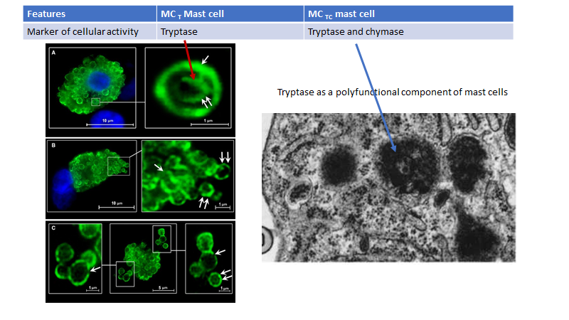
Topics | connective tissue mast cell | mucosal mast cell |
Another name | Also known as MCTC mast cell | Also known as MCT mast cell |
Location | Skin , intestinal submucosa, breast and axillary lymph nodes | Lungs, intestinal mucosa |
Granules and its internal structure | Granules with Lattice like internal structure | Granule with a scroll like internal structure |
Granules contain | Tryptase and chymase | Only tryptase |
Differences between the electrical and chemical synapses.
Differences between the electrical and chemical synapses.
For learning anatomy, please visit :
My youtube channel : @easyhumanatomy73
My website : http://easyhumananatomy.com
My facebook page: https://www.facebook.com/easyhumanatomy/
My blog: http://www.easyhumanatomy73.blogspot.com
My blog: Difference between : http://www.microscopicanatomybd.blogspot.com
Chemical synapses | Electrical synapses |
It is present in higher vertebrates. | It is present in both lower and higher vertebrates and invertebrates. |
Nerve impulse is transmitted using a neurotransmitter. | Nerve impulse is transmitted using ions. |
Unidirectional transmission. | Bi-directional transmission. |
Gaps between cells are around 20 nm | Smaller gaps - only 3 - 5 nm |
Transmission is relatively slow - several milliseconds. | Transmission is fast - almost instant. |
Either inhibitory or excitatory. | Excitatory. |
Signal remains strong. | Signal will disappear over time. |
Sensitive to pH and hypoxia. | Insensitive to pH and hypoxia. |
Vulnerability to fatigue. | Relatively less vulnerable to fatigue. |
Difference between pulsation of jugular vein and carotid artery
Difference between pulsation of jugular vein and carotid artery
For learning anatomy, please visit :
My youtube channel : @easyhumanatomy73
My website : http://easyhumananatomy.com
My facebook page: https://www.facebook.com/easyhumanatomy/
My blog: http://www.easyhumanatomy73.blogspot.com
My blog: Difference between : http://www.microscopicanatomybd.blogspot.com
PULSATION OF JUGULAR VEIN | PULSATION OF CAROTID ARTERY |
No pulsations palpable. | Palpable pulsations. |
Pulsations obliterated by pressure above the clavicle. | Pulsations not obliterated by pressure above the clavicle. |
Level of pulse wave decreased on inspiration; increased on expiration. | No effects of respiration on pulse. |
Usually two pulsations per systole (x and y descents). | One pulsation per systole. |
Prominent descents. | Descents not prominent. |
Pulsations sometimes more prominent with abdominal pressure. | No effect of abdominal pressure on pulsation |
Subscribe to:
Posts (Atom)