Anatomy books

Sunday, January 31, 2016

MS ophthalmology bd: Anatomy brief guide line

Bone :

Base of skull :

Orbit : 

  1. boundary,
  2.  walls, 
  3. content, 
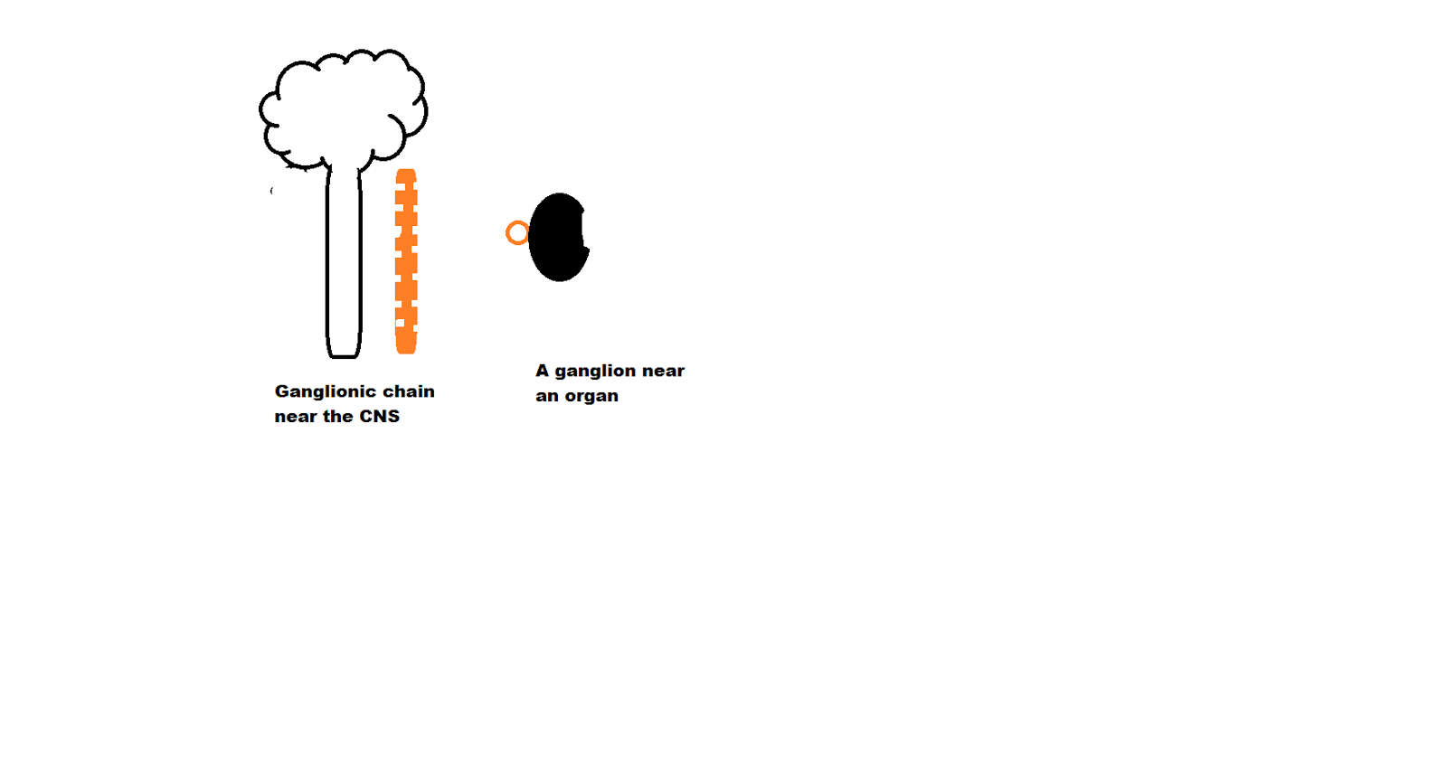
  4. lacrimal gland: type of gland, nerve supply, it is related with which parasympathetic ganglion, it is related with which nucleus
  5. Superior orbital fissure : structures passing through it 
  6. optic canal : structures passing through it 
Frontal bone : orbital surface : important relation
Maxilla

General anatomy: 

Nervous system : 

  1. classification of nervous system
  2. composition of nervous system 
  3. classification of neuron according to polarity, function,
  4. What is nerve?Mention organization of nerve. 
  5. Mention some special features of optic nerve 
  6. classify neuroglia  with functions 

CVS: 

  1. Define circulation. 
  2. Define end artery . Classify end artery with example.
  3. Why central artery of retina is known as true end artery 
  4. define capillary . classify capillary with example
  5. what do you mean by blood retinal barrier 

Muscular tissue : 

  1. Classify muscle histologically 
  2. Mention important features of skeletal , cardiac and smooth muscles 
  3. Mention the types of Extraocular muscle and intraocular muscles
Nervous tissue: 
  1. composition of nervous tissue 
  2. classification of nervous tissue 
  3. myelination in central and peripheral nervous system 
  4. regeneration of peripheral nerve 

General histology : 

  1. Define tissue. Mention the name of basic tissue 
  2. Epithelium : definition, features, classification of epithelium, Classification of covering epithelium(both simple and stratified) , difference between non keratinized and keratinized stratified squmous epithelium, what is the lining epithelium of cornea, conjunctiva and corneal endothelium 
  3. Connective tissue : composition of connective tissue , classify connective tissue proper , sclera : what types of tissue 

General embryology: 

  1.  3rd week of development : development of neural tube 
  2. name of brain vesicle and structure derived from it 
  3. development of lens, cornea , retina , 

Viscera: 

Eyeball : 

anatomical position : 
1. Medial rectus muscle is near the sclero cornea junction 
2. optic nerve lies posterior inferiorly

  1. Cornea: layers of cornea, which layer is thickest, why cornea is transparent, which layer maintain hydration of cornea, how cornea get nutrition , nerve supply of cornea 
  2. Aqueous humor : formation, drainage, clinical anatomy : glucoma 
  3. vascular coat: parts, formation and functions of iris and ciliary body 
  4. retina : development, blood supply, name of neuron present within the retina, how optic nerve is formed ? 
  5. Visual pathway: lesion in optic nerve,optic tract 
  6. what is optic radiation?
  7. what is lateral geniculate body
  8. accommodation reflex 
  9. pupillary light reflex

Brain: 


  1. boundary of occipital lobe, 
  2. show visual area 17, 18, 19 in superolateral surface and medial surface , 
  3. what do you mean by macula sparing ? 
  4. Mention the blood supply of occipital lobe.
  5. parts of diencephalon, 
  6. parts of thalamus , 
  7. what is internal capsule ? 
  8. blood supply of internal capsule. 

Cranial nerve: 


  1. Why trochlear nerve is injured more frequently ? 
  2. how can you diagnose trochlear nerve lesion? 
  3. Facial colliculus is related with which nerve? how can you diagnose injury of that nerve? 
  4. Which colliculus of mid brain is related with vision? 

Lacrimal apparatus : 


  1. mention the name of different parts of it 
  2. how this developed ?
  3. where nasolacrimal duct is open? 
  4. Relation of lacrimal sac.
  5. nerve supply of lacrimal gland 
  6. histological features of lacrimal gland 

Eyelid:


  1. Mention the different layer of it 
  2. how many glands are there in eyelid
  3. Mention the name of muscle present in eyelid 
  4. mention the nerve supply of upper and lower eyelid 

Short questions of thorax

Thorax: 

1. Mention the formation of thoracic cage ?

Ans: Anteriorly, sternum , laterally ribs with costal cartilage(hyaline cartilage) and posteriorly throcic vertebra with intervertebral disc

2. What is thoracic inlet and outlet? 

Ans: upper aperture of thoracic cage and lower aperture of thoracic cage 

3. which structures close the thoracic inlet and outlet?

Ans: The suprapleural membrane covers the thoracic inlet and the diaphragm covers the thoracic outlet. 

4. what is mediastinum? 

Ans: the space between two lungs is known as mediastinum 

5. mention the name of important nerve of thoracic region ?

Ans: Phrenic nerve , intercostal nerve, vagus nerves, recurrent lar;yngeal nerve (branches of vagus nerve) 

6. What do you mean by intercostal nerves? 

Ans: They are ventral rami of upper eleven thoracic spinal nerves 

7. What is the name of ventral ramus of 12th  thoracic spinal nerve?

Ans: Subcostal nerve 

8. Posterior relation of manubrium sternae? 


Ans: lower half arch of aorta and upper half branches of arch of aorta( brachiocephalic trunk, left common carotid and left subclavian artery) and left brachiocephalic vein

9. how sternocostal surface of heart is formed?  

Ans : this surface is formed by four chamber of heart
  1. 2/3 right ventricle
  2. 1/3 left ventricle
  3. right atrium and auricle 
  4. left auricle of left atrium 
10. how diaphragmatic surface of heart is formed?
Ans: 1/3 of right ventricle and 2/3 of left ventricles 

11. How base of heart is formed ?

Ans : By two atria, 2/3 by left atrium (because most of the part of left atrium is situated posteriorly )
and 1/3 of right atria (most of the part right atria is situated anteriorly)
  
Q: mention the anatomical point of heart 
Ans : this is the viscera of heart with root of the great vessels with pericardium (if present )
a) apex is directed downward, forward and to the left 
b) base is directed backward and to the right 
c) sternocostal surface of heart is directed anteriorly and the diaphragmatic surface of heart is directed inferiorly 
 Q: 


Monday, January 18, 2016

Ordinary connective tissue (connective tissue proper)

Ordinary connective tissue (connective tissue proper)

This types of connective tissue bears no special character or features.They only connect two types of tissue together. For example transitional epithelium of urinary bladder connected with smooth muscles layer by connective tissue known as lamina propria. 

Classification of ordinary connective tissue:

1. Loose connective tissue : why it is known as loose because it contain loosely arranged connective tissue fibres 
2. Dense connective tissue : why dense ? because it contain densely arranged connective tissue fibres. According to manner of arrangement of connective tissue fibres it is again two types:
Dense irregular connective tissue: dense connective tissue fibres arranged irregularly 
Dense regular connective tissue : dense connective tissue fibres arranged regularly   


Loose connective tissue contain large number of connective tissue cells but less and loosely arranged connective tissue fibres. Example: lamina propria of mucosa of intestine (it handle stress and allow distension)  


Dense irregular connective tissue : contain less connective tissue cells but densely and irregularly arranged connective tissue fibres: Example : dermis of skin ( irregularly arranged connective tissue fibres bear more stress from any direction ) 

Dense regular connective tissue contain less connective tissue cells but densely and regularly arranged connective tissue fibres  : example: tendon (it bear more stress but from particular direction ) 

Thursday, January 14, 2016

Ganglia and nucleus : similarity and difference

Nucleus : 

collection of nerve cells bodies within the central nervous system.

SN:

  1. Big collection of nerve cells body within the central nervous system is known as grey matter. 
  2. Small collection of nerve cells body within the central nervous system is known as nucleus. 
  3. Some time part of grey matter is also known as nucleus. 
  4. Some time the axons of nucleus leaves the central nervous system as peripheral nerve

Ganglia : 

Collection of nerve cells bodies outside the central nervous system is known as ganglia.
SN:
Nerve cells bodies of ganglia some time synapses with axon of other neuron 


Some time axon of other neuron only passage through the ganglia 


Some time ganglia connect with each other so axon of one ganglia passage above ganglia or below ganglia 


Some ganglia near to central nervous system but some are nearer to target organ 

Similarity between nucleus and ganglia : both are collection of nerve cells bodies 

Difference between nucleus and ganglia: nucleus lies within CNS and ganglia lies within PNS  

Wednesday, January 13, 2016

Nucleus: at a glance

Nucleus 

Parts of nucleus
      Nuclear membrane
      Nucleolus
      Chromatin &  nucleoplasm
Nuclear membrane
      It is a double layer membrane. The outer layer of membrane attached with ribosomes and continuous with rER.

      Pores present 

Nucleolus:  
1. It is a structure found in the nucleus of cells.
2. It is formed by proteins and nucleic acids. 
3. It is occupies about 25% of the volume of the cell nucleus.

Function:

 It transcribes ribosomal RN(rRNA)
The synthesis of RNA is regulated by five different chromosomes (13, 14, 15, 21, and 22). 

 Malfunction of nucleoli : 

mutation causes malfunction  of nucleus and it is related to following disease like Weine syndrome, Treacher Collins Syndrome and dyskeratosis.
      Chromatin / Chromatin granule

        The readily stainable substance of a non dividing cell nucleus consisting of DNA and RNA and various     
            proteins; during mitotic division it condenses into chromosomes
      They are two types: 

Heterochromatic
Densely packed portion of chromatin which is genetically inactive.
Bluish in colour. 

Euchromatic
 
uncoiled portion of chromatin which is genetically active.
 Lightly stain 

What is chromosome?
      it is  a  threadlike structures, which visible during cell division and is composed by
      DNA,
      Small RNA,
       histone and non-histone protein. (consisting of genes that are responsible for the transmission of hereditary characteristics)

Types of chromosome:
 Autosomes chromosome:
      The genes which regulate body character.
      They are 22 pairs
 Sex chromosome
      The genes which regulate sex character.
      They are 1 pair
      In male one X and one Y .
                  XY
      In female two X.  
                        XX
Chromosomes number in human body
      46
      /23 pairs
       22 pairs autosomes + 1pair sex chromosome
Diploid: A cell or an organism consisting of two sets of chromosomes:
Usually, one set from the mother and another set from the father.
In a diploid state, the haploid number is doubled, thus,
This condition is also known as 2n.
Haploid: The number of chromosomes in a gamete of an organism, symbolized by n

Parts of a chromosome
Each chromosome has a constriction point called the centromere, which divides the chromosome into two sections, or “arms.”
 The short arm of the chromosome is labeled the “p arm.”
The long arm of the chromosome is labeled the “q arm.
Chromatid - one of two identical strands into which a chromosome splits during mitosis


Tuesday, January 12, 2016

Short question on Superior extremity

1. How many compartment present in arm?

Ans : Anterior compartment : which is innervated by musculocutaneous nerve
       Posterior compartment : which is innervated by radial nerve

2. How many compartment present in forearm?

Ans: Anterior compartment : which is innervated by mainly by median nerve and partly by ulnar  nerve
       Posterior compartment : which is innervated by radial nerve

3. Which nerve of upper limb innervated two compartment of upper limb?

Ans : Radial nerve which is innervated both posterior compartment of arm and forearm

4. Musculocutaneous nerve, what is the meaning of musculocutaneous ?

Ans: This nerve pierce the corabobrachialis muscle and end as cutaneous nerve known as lateral cutaneous nerve of forearm.

5. Which nerves innervated muscles of hand ?

Ans: Mainly ulnar nerves supply most of the muscles of hand  and some are supplied by median nerve

6. Which nerve plexus supply upper limb? 

Ans: Brachial plexus

7. What types of plexus is it ?

Ans: somatic plexus

8. How it is formed? 

Ans: it is formed by ventral rami of C 5 to 6 and T 1 spinal nerve

9. What do you mean by pre- fixed and post fix types of brachial plexus 

Ans: pre- fixed brachial plexus : formed by C 4 to 8 
Post fixed: formed by C6 to 8 T1-2

10 What is nerve plexus?

Ans: net of nerves. they are ventral rami of spinal nerves at different label


Monday, January 11, 2016

Thymus : Histological identifying points with easy drawing idea

Thymus : it is a primary lymphatic organ.

Features of Thymus:

  1. It is covered by capsule 
  2. trabeculae, the extension of capsule divide the organ into lobules 
  3. Each lobule has outer cortex and inner medulla 
  4. it contain no lymphatic nodule 
  5. It contain only T lymphocytes 


Histological features of Thymus
1. each thymic lobule has outer cortex and inner medulla
2 inner medulla contain Hassel's corpuscle
3. outer cortex contain no lymphatic nodule

Sunday, January 10, 2016

Lymph node : Histological features with easy drawing idea

Lymph node: 

  1. It is the one of the important lymphatic organ. 
  2. It is a secondary lymphatic organ 
  3. It has one efferent lymph vessel but many afferent lymphatic vessel 
  4. Its efferent lymphatic vessel leave through hilum 
  5. Its afferent lymphatic vessels enter to this organ at different location 
  6. It is covered by capsule
  7. It has two part outer cortex and inner medulla 
  8. Outer cortex contain lymphatic nodule 

Lymphatic nodule: 

A round shape structure, which is formed by aggregation of lymphocytes
It is darkly stain (deep blue) because lymphocyte contain big nucleus with little cytoplasm

Which lymphatic organ contain lymphatic nodules?

Ans:  
  1. Lymph node 
  2. Spleen 
  3. Tonsil 
GULT: Gut associate lymphatic tissue contain aggregation of lymphocyte mainly in initial and terminal part of alimentary tract 
BULT: Bronchus associate lymphatic tissue contain aggregation of lymphocyte at initial part of respiratory system 

Germinal center: 

A pale stain area within the lymphatic node 
It contains B Lymphocyte 

Why this area pale ? 

It contain immature lymphocyte 
the cytoplasm of immature lymphocyte is bigger the nucleus and the number of cells are less so this area seem to paler than periphery 

 Special feature for identification of histological slide :

  1. Subscapsular sinus is present ( unique feature of Lymph node) 
  2. It has two parts outer cortex and inner medulla 
  3. Outer cortex contain lymphatic nodule 


Friday, January 8, 2016

Organization of peripheral nerve Vs organization of skeletal muscle: easy understanding

Peripheral nerve : it is a collection of nerve bundles surrounded by a connective tissue coat, epineurium . 


Nerve bundle : it is a collection of nerve fibres surrounded by a connective tissue coat, perineurium 


Nerve fibre: it is a process of a neuron (usually axon) surrounded by myeline sheath (schwann cells) and  a connective tissue coat, endoneurium. 


Skeletal muscle: it is a collection of muscle bundles surrounded by a connective tissue coat, epimysium.


Muscle bundle: it is a collection of muscle fibres (muscle cells) surrounded by a connective tissue coat, perimysium.


Muscles fibre: when an individual muscle cells are surrounded by a connective tissue coat, endomysium.