Anatomy books

Tuesday, December 24, 2019

Cleavage in first week of development


Cleavage
It is a series of mitotic divisions of the zygote.
Cytoplasm of zygote is successively cleavage (partitioned  ) to form a blastula consisting of increasingly smaller blastomeres (2-cell, 4-cell, 8-cell and so on)
Blastomeres are considered totipotent (capable of forming a complete embryo) upto the 4-8 cell stage (important when considering monozygotic twinning )
Blastomeres form a morula by undergoing compaction that is tight junctions are formed between the cells in the outer cell mass , thereby sealing off the inner cell mass . uvomorulin a glycoprotein found on the surface of blastomeres is involved in compaction

<script data-ad-client="ca-pub-3738618711723990" async src="https://pagead2.googlesyndication.com/pagead/js/adsbygoogle.js"></script>

Monday, December 23, 2019

The diaphragms in the body

The diaphragms in the body are
1. diaphragma sellae
2. oral diaphragm (mylohyoid muscle)
3.supra pleural membrane (sibson's fascia )
4.the diaphragm
5. pelvic diaphragm
6. urogenital diaphragm
<script data-ad-client="ca-pub-3738618711723990" async src="https://pagead2.googlesyndication.com/pagead/js/adsbygoogle.js"></script>

Saturday, December 21, 2019

Summary of pericardial sinuses both transverse sinus and oblique sinus

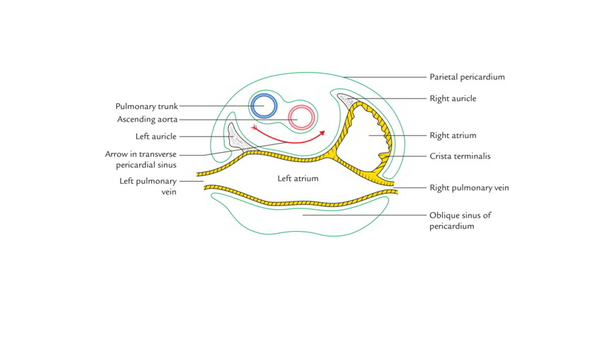
Sinuses of pericardium
Pericardial sinuses are potential space within the serous pericardiam .
There are two pericardial sinuses: transverse and oblique.
Oblique sinus
Definition :
It is the cul-de-sac behind the left atrium and is closed on all sides except below .
It is placed between parietal and visceral layer of pericardium
<script data-ad-client="ca-pub-3738618711723990" async src="https://pagead2.googlesyndication.com/pagead/js/adsbygoogle.js"></script>
 Shape:
 Inverted ‘J’ shaped
Boundaries:
Anteriorly : left atrium and visceral layer of serous pericardium
Posteriorly :  parietal layer of pericardium and fibrous pericardium
Right side : right pair of pulmonary veins and inferior vena cava
Left side : left pair of pulmonary veins
Above : upper margin of left atrium
Inferiorly : open
Special note:
The roof of oblique sinus and floor of transverse sinus is separated by upper margin of
the left atrium only , along which a bilaminar fold of serous pericardium extends from the
upper right to the upper left pulmonary veins
Development:
It develops as an effect of absorption of 4 pulmonary veins into the left atrium.
Function of oblique sinus:
 The oblique sinus permits the distension of left atrium during return of oxygenated
blood in it from the lungs.
Transverse sinus
Definition :
It is a transverse passage between two tubular reflections of serous pericardium and is lined by visceral layer only. It is an inter-visceral space.
Boundaries :
In front : ascending aorta and pulmonary trunk enclosed in a single tube of serous pericardium because both are developed from the truncus arteriosus
Behind intra-pericardial part of superior vena cava and upper margin of left atrium
Above bifurcation of pulmonary trunk
Below upper surface of left atrium
Development:
it is developed after degeneration of the central cells of the dorsal mesocardium .


Summary of transverse sinus of pericardium with clinical anatomy


Transverse sinus
Definition :
It is a transverse passage between two tubular reflections of serous pericardium and is lined by visceral layer only. It is an inter-visceral space.
Boundaries :
In front : ascending aorta and pulmonary trunk enclosed in a single tube of serous pericardium because both are developed from the truncus arteriosus

<script data-ad-client="ca-pub-3738618711723990" async src="https://pagead2.googlesyndication.com/pagead/js/adsbygoogle.js"></script>
Behind intra-pericardial part of superior vena cava and upper margin of left atrium
Above bifurcation of pulmonary trunk
Below upper surface of left atrium
Development:
it is developed after degeneration of the central cells of the dorsal mesocardium .

Summary of oblique sinus of pericardium with clinical anatomy

Oblique sinus
Definition :
It is the cul-de-sac behind the left atrium and is closed on all sides except below .
It is placed between parietal and visceral layer of pericardium


 Shape:
 Inverted ‘J’ shaped

Boundaries:
Anteriorly : left atrium and visceral layer of serous pericardium
Posteriorly :  parietal layer of pericardium and fibrous pericardium
Right side : right pair of pulmonary veins and inferior vena cava
Left side : left pair of pulmonary veins
Above : upper margin of left atrium
Inferiorly : open

Special note:
The roof of oblique sinus and floor of transverse sinus is separated by upper margin of
the left atrium only , along which a bilaminar fold of serous pericardium extends from the
upper right to the upper left pulmonary veins

Development:
It develops as an effect of absorption of 4 pulmonary veins into the left atrium.
Function of oblique sinus:
 The oblique sinus permits the distension of left atrium during return of oxygenated
blood in it from the lungs.

Tuesday, December 17, 2019

Summary of Hepato-renal recess/ Pouch of Morison


Hepato-renal recess/ Pouch of Morison
Definition:
The hepatorenal recess[1] (subhepatic recesspouch of Morison or Morison's pouch) is the space that separates the liver from the right kidney. As a potential space, the recess is not filled with fluid under normal conditions.

<script data-ad-client="ca-pub-3738618711723990" async src="https://pagead2.googlesyndication.com/pagead/js/adsbygoogle.js"></script>


Boundaries :
Anteriorly :
·         inferior surface of right lobe of liver and gall bladder
Posteriorly :
·         right suprarenal gland
·         Upper part of right kidney
·         2nd part of duodenum
·         Hepatic flexure of colon
·         Transverse mesocolon
Superiorly
·         Inferior layer of coronary ligament
Inferiorly
·         Opens into general peritoneal cavity

Clinical anatomy :
 However, fluid can collect in this space in circumstances where the abdomen fills with fluid, such as mostly ascites and hemo peritoneum. This fluid may be seen on ultrasound or computed tomography (CT scan).


 The intraperitoneal fluid, be it blood, ascites, or dialysis, collects in this space and may be visualized, most commonly via ultrasound or computed tomography (CT) scanning. As little as 30 or 40 ml of fluid in the abdominal cavity may be visualized in this space.
Early visualization of fluid in the hepatorenal recess on FAST scan may be an indication for urgent laparotomy