Para-follicular cells of thyroid gland
|
Topics
|
Para-follicular cells
|
|
Secretion
|
Calcitonin
|
|
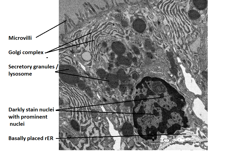
Shape and size of cell
|
It is larger than the follicular cell and the shape of the
cell is Round
|
|
Staining
|
It takes less staining
|
|
Exposure to luminal surface
|
It is never exposed to luminal surface, it is located
peripheral part of thyroid follicle
|
|
Nuclei
|
It contain large pale stain nuclei
|
|
Golgi complex
|
Large amount of golgi
complex are present but no definite
location
|
|
Secretory vesicles
|
It contains numerous secretory vesicle present though out
of the cell
|
|
rER
|
Present in small amount but no definite location
|
|
Lysosome
|
Present but not abundant
|
|
Endocytotic
vesicles or colloidal resorption droplets
|
No present
|
|
Microvilli
|
Absent
|
|
Development
|
4th pharyngeal pouch
|
|
Derived from
|
Neural crest
|