How to identify stratifed cuboidal epithelium in histological slide
1. only 2-3 cells layers are present
2. name of stratified epithelium are decided according to the shape of the surface cell
Ex: ducts of sweat glands

How to identify stratifed cuboidal epithelium in histological slide
1. only 2-3 cells layers are present
2. name of stratified epithelium are decided according to the shape of the surface cell
Ex: ducts of sweat glands
How to identify pseudostratified ciliated columnar epithelium
For learning anatomy, please visit :
My youtube channel : @easyhumanatomy73
My website : http://easyhumananatomy.com
My facebook page: https://www.facebook.com/easyhumanatomy/
My blog: http://www.easyhumanatomy73.blogspot.com
My blog: Difference between : http://www.microscopicanatomybd.blogspot.com
1. single layer of tall and short columnar shape ( height of some cells are more and height of some cells are less but all cells rest on basement membrane )cells rest are on basement membrane
2. cell contain single centrally place oval nuclei , located near the basal region
3. Level of nuclei are different so it is look like stratified epithelium but all cells are rest on basement membrane . it stratified epithelium only basally place cells layer rest on basement membrane . other cells layer rest on previous cell layer .
Ex: lining epi of trachea, bronchi, epididymis, vas deferens

How to identify simple columnar epithelium in histological slide
1. single layer of columnar shape ( height more and width less)cells are rest on basement membrane
2. cell contain single centrally place oval nuclei , located near the basal region
ex: lining epi. of stomach, lining epi. of gall bladder

How to identify simple cuboidal epithelium in histological slide
1. single layer of cuboidal shape (same height and width) cells are rest on basement membrane
2. cell contain single centrally place round nuclei
ex: thyroid follicles, kidney tubules

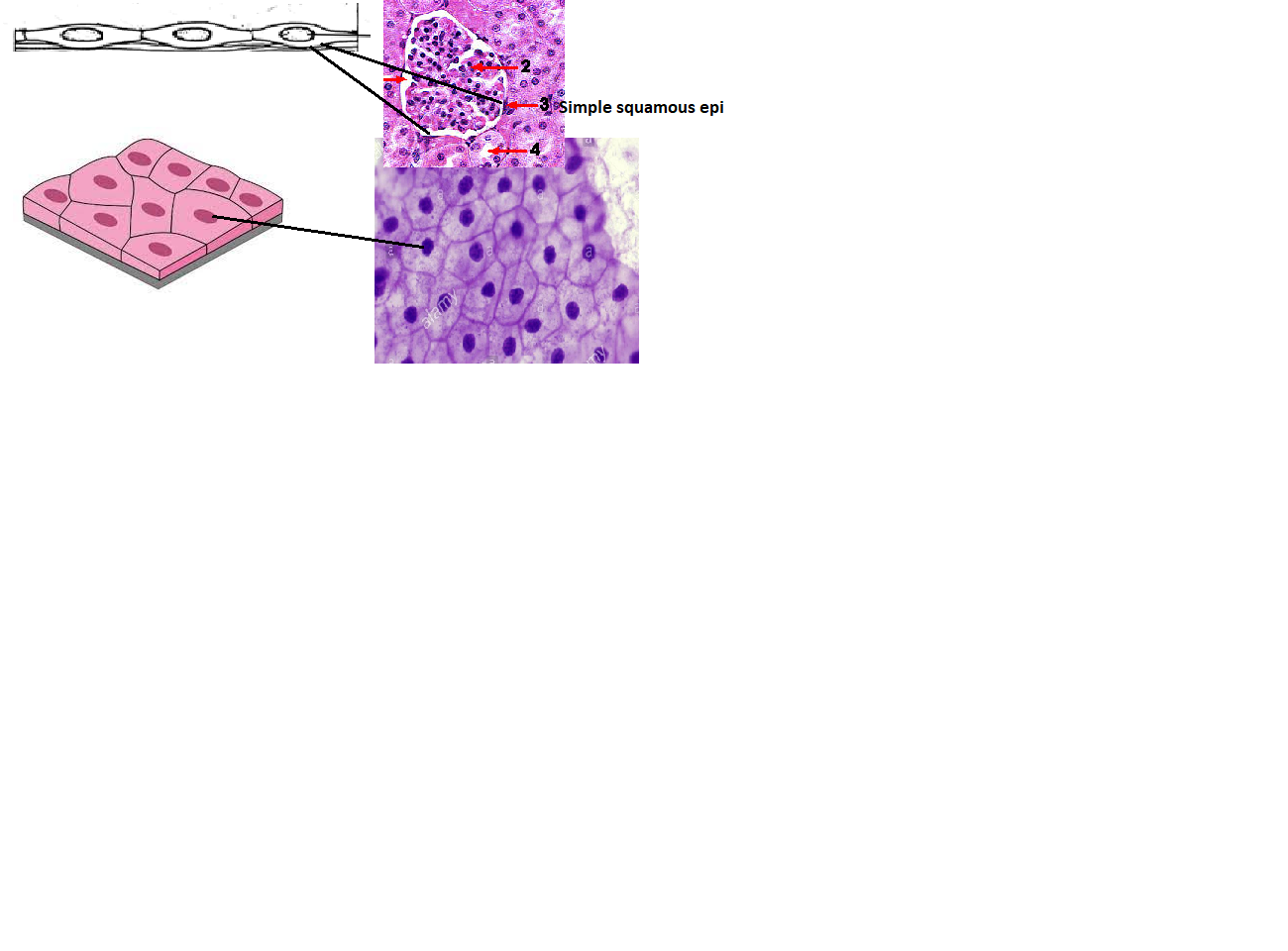
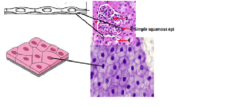
How to identify Simple squamous epithelium in histological slide
1. single layer of flattened cells are rest on basement membrane
2. cell contain single centrally place flattened oval nuclei
ex: endothelium, mesothelium, endocardium