Fate of cloaca
What is cloaca ?
Cloaca : part of hindgut (derived
from endoderm) caudal to attachment of allantois, which is common chamber for
hindgut & urinary system


It is divided into two parts ventral and dorsal by urorectal septum
ventral part is called primitive urogenital sinus
dorsal part is called primitive rectum
ventral part is called primitive urogenital sinus
dorsal part is called primitive rectum
What are the parts of
primitive urogenital sinus ?
Vesico-urethral canal and definitive urogenital sinus
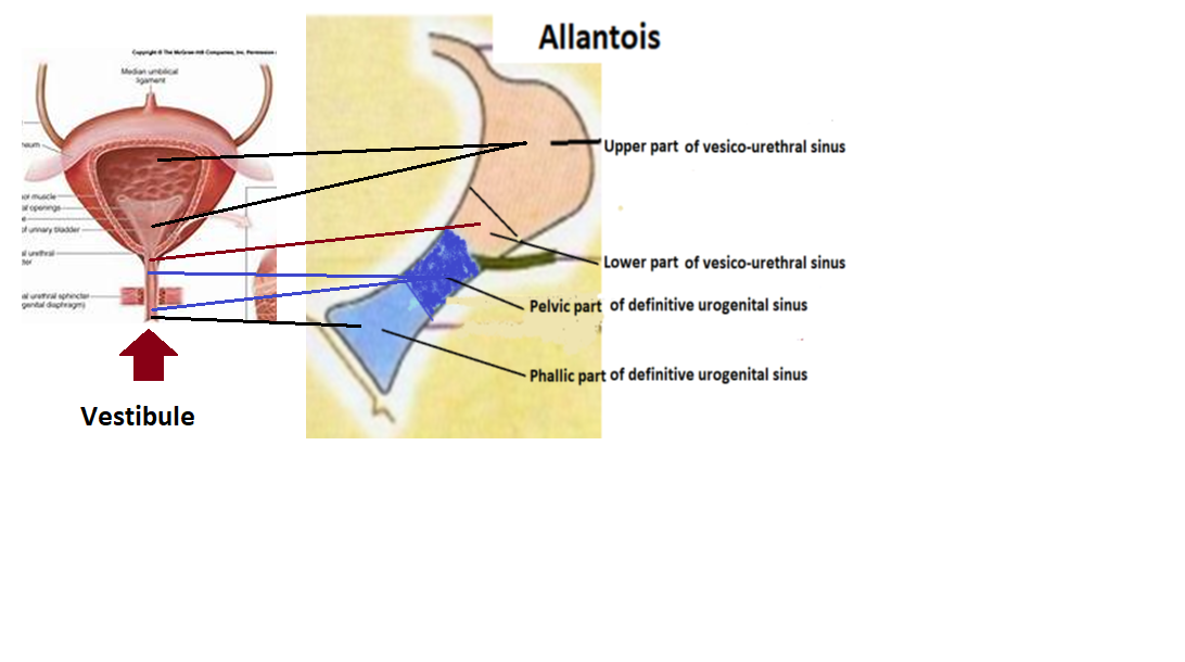
From above down ward different parts of primitive urogenital sinus
1. Upper part of vesico-urethral canal
2. Lower part of vesico-urethral canal
3. Pelvic part of definitive urogenital sinus
4. Phallic part of definitive urogenital sinus
Cloaca
Rectum & upper part of anal canal
Upper part of primitive urogenital sinus is
known as
Vesico-urethral canal : it has 2 part
Upper part of vesico-urethral canal develops urinary bladder
Lower part of vesico-urethral canal upper part of prostatic
urethra of male & most of the proximal part of female urethra
Lower part of primitive urogenital sinus is
known as
Definitive urogenital sinus: it has 2 part
Pelvic part of definitive urogenital sinus: prostaic urethra &
membrous urethra in male and lower small part of female urethra
Phallic part of definitive urogenital sinus: penile part of male urethra &
terminal part of female urethra which open into the vestibule of valva
Cloacal malformation : In rare cases (1 in every 50,000 babies), this
process does not work properly and these tracts do not separate from one
another completely. A female is said to have developed a “persistent cloaca”
when these three tracts open into one common cavity, with one opening from the
body.
The hind-gut
is at first prolonged backward into the body-stalk as the tube of the allantois;
but, with the growth and flexure of the tail-end of the embryo, the body-stalk,
with its contained allantoic tube, is carried forward to the ventral aspect of
the body, and consequently a bend is formed at the junction of the hind-gut and
allantois.
This bend
becomes dilated into a pouch, which constitutes the endodermal cloaca; into its dorsal part the hind-gut opens, and
from its ventral part the allantois passes forward.
At a later
stage the Wolffian duct and Müllerian duct open into its ventral portion.
The cloaca
is, for a time, shut off from the anterior by the cloacal membrane, formed by
the apposition of the ectoderm and endoderm, and reaching, at first, as far
forward as the future umbilicus.
Behind the
umbilicus, however, the mesoderm subsequently extends to form the lower part of
the abdominal wall and pubic symphysis.
By the growth of the surrounding tissues the cloacal membrane comes to lie at the bottom of a depression, which is lined by ectoderm and named the ectodermal cloaca.


No comments:
Post a Comment