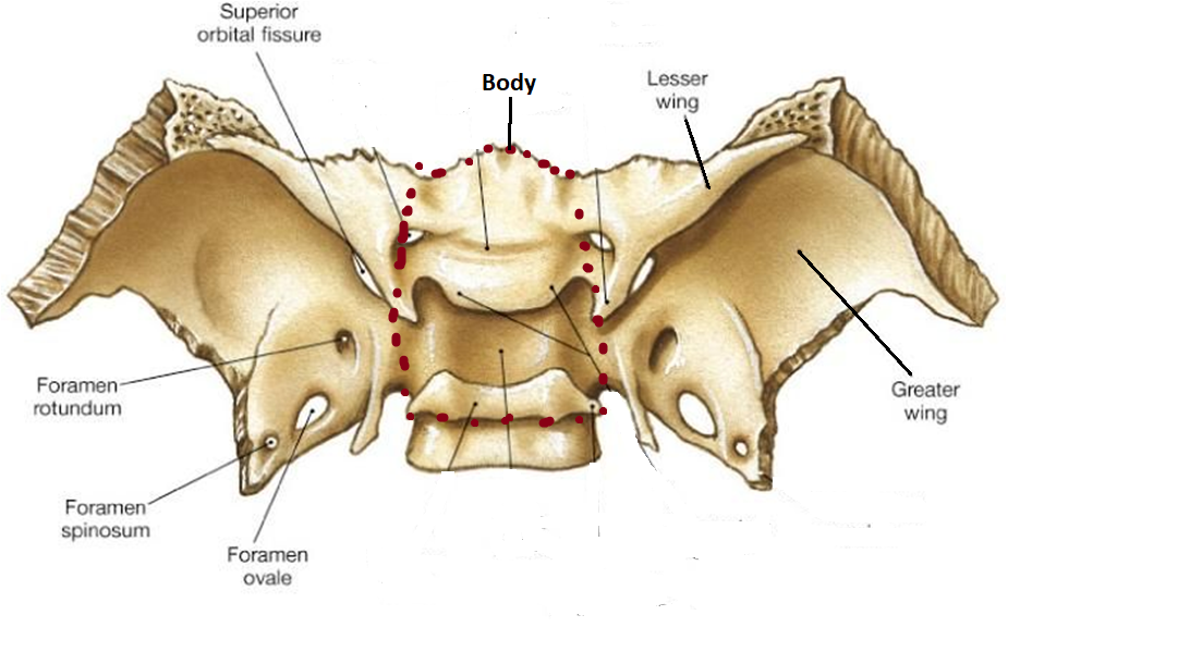
Anatomy of sphenoid
Anatomical points
- Body lies centrally
- superior surface of body lies horizontally
- Two greater wings & two lesser wings project laterally from the sides of the body
- Superior surfaces of greater wings are deeply concave
- Two pterygoid processes are directed downwards from adjoining parts of the body and greater wings Morphological type of bone: pneumatic irregular bone
https://www.amazon.com/gp/product/B08L7R9DL4/ref=as_li_tl?ie=UTF8&camp=1789&creative=9325&creativeASIN=B08L7R9DL4&linkCode=as2&tag=ezhumanatomy-20&linkId=e4dff7c8da8662c897ebe16b667f67f1
Parts of sphenoid bone
Body
Two greater wings
Two lesser wings
Two pterygoid processes
Body: It has six surfaces : superior , inferior , anterior , posterior and two lateral surfaces
Shape : cuboid
It contains two large air sinuses which are separated from each other by a septum
Superior or cerebral surface
Articulates with ethmoid bone anteriorly and basilar part of occipital bone posteriorly. It shows:
Ethmoidal spine : it is articulate with posterior border of cribriform plate of ethmoid bon
Jugum sphenoidale: smooth area which is the part of anterior cranial fossa . this part is related with gyri recti of cerebrum and olfactory tracts
Sulcus chiasmaticus: it is a groove which connects both optic canal . it lodges optic chiasm
Tuberculum sellae: round elevation it bears middle clinoid processe
Sella turcica: deep depression behind the tuberculum sellae . deepest part of sella turcica is
known as hypophyseal fossa
Dorsum sellae: square shape bone which form posterior boundary of sella turcica . it bear
posterior clinoid processes
Clivus: slopping area behind dorsum sellae . it continue with basilar part of occipital bone this part is related with pons
- Inferior surface
Rostrum of sphenoid
Sphenoidal conchae
Vaginal processes of medial pterygoid plate
Anterior surface
Sphenoidal crest articulates with the perpendicular plate of ethmoid leading to formation of a part of the septum of nose.
Posterior surface: Basilar part of occipital bone
Lateral surface
- It is united with the greater wing and medial pterygoid plate ,
Carotid sulcus is present in the lateral surface which is extend from superior orbital fissure to foramen lacerum s lodging cavernous sinus and internal carotid - Development:
- up to rostral half of sella turcica is developed from neural crest
- caudal half of sella turcica is developed from paraxial mesoderm


No comments:
Post a Comment